Art of Medicine
May 2010
American culture places great emphasis on body shape. There is a widely held presumption that “diet plus exercise = looking good.” This premise gives rise to huge expenditures of time and resources in all-too-often frustrating attempts to “get in shape,” but what is not considered is that shape and size may have entirely different biological underpinnings. Many individuals successfully lose weight and significantly reduce body size, only to remain unhappy with their residual shapes.
To a certain degree size may be under one’s control by the intentional modulation of caloric intake and burn, but there is little data to support the idea that even the most stringent efforts can effectively and permanentlychange body configuration or fat distribution. For example, dietary manipulation can affect overall size, but though it may temporarily shrink both waist and hips, it will not necessarily bring about the desired waist-to-hip ratio. Because many Americans believe that body shape can be controlled by behavior, societal judgment is often levied against patients who choose to manipulate their native body contours surgically. Absent evidence that shape is inborn, many continue to struggle for decades, only to fail to reach their goals. Since surgery requested later in life is more complex, and complication rates can be higher, this misconception has ethical implications.
Anatomy and body shape are evaluated routinely by a host of imaging techniques. Medical imaging underwent major expansion in the late twentieth century with the introduction of CAT scan technology, magnetic resonance imaging, and other computer-based modalities. During those same years, however, the advent of digital cameras resulted in a shift in clinical photography to a less scientific “point-and-shoot” mentality, which produced an explosion of case-related patient images that were often published with no consistent standardization of technique. The outcomes of plastic surgery intervention are often evaluated by looking at these less-than-ideal “before and after” snapshots.
Such documentation fails to provide accurate and quantifiable data to support the notion that surgery has effected permanent change, or that the underlying condition could only be changed by surgery in the first place. Fortunately for our future understanding of this complex issue, standardized imaging technologies and software now exist to address long-unanswered questions about the inheritance of body shape and the quantification of surgical results. A new and unique monozygotic mirror-twin model incorporating standardized photographic techniques provides a tool for investigating questions of anatomic development and adult human form.
Facial skin features historically were thought to stem from a combination of genetic and environmental influences. In the past, to help determine the genetic origin of a facial skin feature, correspondence of surface findings was erroneously sought by comparing the same sides of two twins [1]. More recently, I have used highly standardized photographic techniques and skin surface analysis to address questions of inheritance of anatomic features [2]. With technical insight from Kalev Peekna, I developed a formal digital method to easily account for the phenomenon of mirroring in twins which, though previously ill-defined by science, has been long acknowledged among twins themselves. Anatomic mirroring is the term used to describe the phenomenon that a lesion or anatomic structure on one side of a monozygotic (MZ) twin is found in a similar location on the opposite side of the co-twin (e.g., a mole on twin A’s right cheek can be paired to a mole on twin B’s left cheek). Our technique was therefore developed to definitively and reproducibly diagnose mirroring and allow for its differentiation from simple same-side concordance in order to show the genetic contribution to facial shape [3].
Figure 1 shows typical concordance of skin features in a pair of MZ twins who exhibit no anatomic mirroring. Correspondence in the skin surface findings in another set of twins can only be appreciated if opposite sides of the face are carefully examined (figure 2).
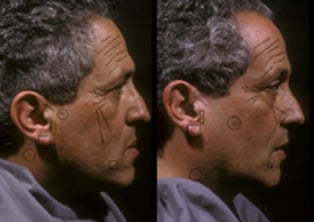
Figure 1. (Click the magnifier to enlarge image.) Detailed analysis of the same sides of the faces of two concordant, non-mirrored MZ twins reveals striking similarities. These similarities include the same number and configuration of wrinkle creases on both the forehead and brow, nearly identical crow’s feet wrinkle lines with similar branching patterns at the corners of the eyes, similar helical root creases, pre-tragal creases, identical oblique earlobe creases, and a series of skin lesions that appear to have migrated at different rates during early embryonic development, with each feature being more anterior in twin B. None of the findings present on the right sides of the twin faces are present on the left.
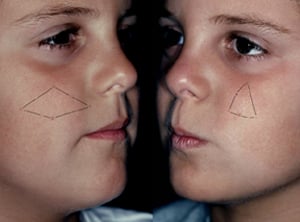
Figure 2. (Click the magnifier to enlarge image.) Both twin A (left) and twin B (right) exhibit a polygon of nevi only on opposite cheeks. It is likely that different rates of embryologic tissue transit account for the slight differences in the shapes of the polygonal arrangement in each twin, although both clusters remain within the boundaries of the anatomic region innervated by the second branch of the trigeminal nerve.
New digital addition and subtraction techniques used to analyze highly standardized images of twins can be employed to study facial shape for the presence of anatomic mirroring [3]. As in radiological techniques used for digital subtraction angiography, images of twin faces are overlapped and then digitally subtracted from each other to determine whether anatomic shape was concordant (present on the same side in both twins) or mirrored (present on the right side in one and left side in the other), as are the twins in figures 3 and 4. Analysis of 27 pairs of monozygotic twins showed that 64 percent of male pairs and 23 percent of female pairs exhibited the mirror phenomenon, and that there was no relationship between the mirror phenomenon and the timing of the first split of the egg in either gender [4]. When the appropriate side of the face was analyzed (i.e., either the same or opposite) in these same twins, nearly 100 percent of skin features were found to be present in both twins [5]. In light of these observations, all future studies of anatomic inheritance should control or consider the mirror phenomenon.
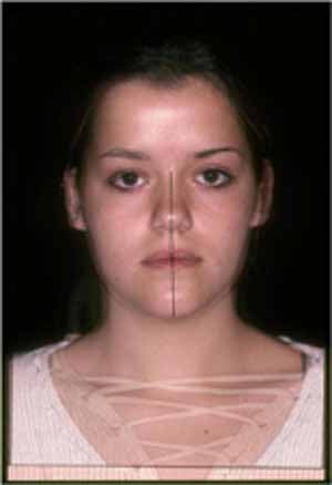
Figure 3. (Click the magnifier to enlarge image.) Representative pair of female mirror twins. Twin A and twin B have been digitally overlapped.
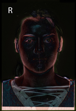
Figure 4. (Click the magnifier to enlarge image.) When digitally subtracted from each other, the images from figure 3 show symmetrical “ghosting” consistent with anatomic mirroring of the pair’s skin findings. (Digital subtraction of the images of concordant twins results in an asymmetrical “ghost,” indicating that the inherent asymmetries of the face are concordant and not mirrored.)
In addition, and perhaps more importantly, the above findings bring the role of environmental influence into question. It is illogical to think that random environmental influence could consistently affect only one side of one twin and only one side (for example, just the mirror-opposite side) of another twin in exactly the same way over their entire lifetimes—whether they were raised in the same or different environments. As a result, environmental influence can be eliminated as a variable if mirroring is analyzed and controlled in the twin study population.
Standardized imaging and digital analysis have preliminarily confirmed the presence or absence of mirroring of body form in MZ twin torsos. Figure 5 illustrates the extreme alignment of anatomy when two concordant male MZ twin torsos are digitally added to each other, but the alignment is lost when the photograph of Twin B is horizontally flipped. Digital subtraction has successfully identified concordance or mirroring in all pairs studied to date. It follows that the body shapes of the twin pairs must be inherently similar (concordant) or similar-but-mirrored, regardless of differences in size [5].
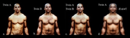
Figure 5. (Click the magnifier to enlarge image.) Left to right: The native state of twin A; the native state of twin B; the digital addition of twin A imposed on twin B, showing near-complete anatomic alignment of the torsos; and, finally, the digital addition of the native state of twin A added to the horizontally flipped image of twin B, showing a dramatic decrease in alignment consistent with a non-mirrored native state.
The same standardized imaging techniques can also be used to accurately quantify postsurgery results, because photographic variance has been nearly eliminated. In figure 6, the postoperative result has been digitally subtracted from the preoperative baseline anatomic state, providing evidence of shape change which can actually be measured. The same methods could be used to track disease progression (e.g. Cushing disease or HIV-related lipodystrophy), the effects of therapeutic interventions, or changes in body configuration due to aging.
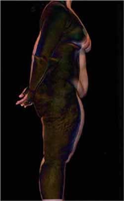
Figure 6. (Click the magnifier to enlarge image.) Standardized digital subtraction analysis (preoperative minus postoperative views) of the surgically imposed shape changes following full-body circumferential reproportioning. This surgery was preceded by weight loss of more than 100 pounds, which had reduced the patient’s size, but had not achieved the patient’s desired shape.
The above findings, developed using a MZ twin approach that controls for the “mirror twin” phenomenon, supports the concept that body surface features and body shape are genetically predetermined. Diet and exercise appear to be able to temporarily alter size, but it seems that only surgery, disease, or trauma can permanently alter shape.
This observation has direct implications for twins and non-twins alike who have concerns about skin or body features. Patients who request body contour surgery (the elective alteration of baseline anatomic form) are often counseled to make lifestyle changes to alter their weight (with the presumption that it will change their shape) before surgery is performed. In light of the findings presented above, patients should instead be counseled to adopt healthy diets and exercise routines that can be maintained throughout adulthood, regardless of the effect on weight preoperatively. Surgery should proceed once metabolic steady state is reached and body weight has stabilized, after several months, so the patient can enjoy an improved body configuration without struggling to maintain an unrealistic daily routine. Data on the genetic inheritance of undesired body shapes could help inform future ethical decisions regarding elective surgery.
The broader implication of these photographic and anatomic findings is that the very structure of the “nature vs. nurture” debate as it pertains to body shape must be reconsidered. It is clear that there may be limits to the effect of environment on anatomic shape.
Gedda L. Twins in History and Science. Springfield, IL: Charles C. Thomas; 1961.
Teplica D, Keith D. A study of the mirror symmetry phenomenon in the faces of 100 sets of monozygotic twins, using rigidly standardized photographic techniques and digital analysis. Paper presented at: 10th International Workshop on Multiple Pregnancy; September 5-7, 1996; Zakopane, Poland. Arch Perinat Med. 1996:1(2).
Teplica D, Peekna K. The mirror phenomenon in monozygotic twins. In: Blickstein I, Keith L. Multiple Pregnancy: Epidemiology, Gestation, and Perinatal Outcome. 2nd ed. London: Taylor and Francis; 2005: 277-288.
Teplica D, Derom C, Peekna K, Derom R. Embryological timing in mirror-image twinning [abstract]. Twin Res Hum Genetics. 2007;10 Suppl:54.
Teplica D. Iconography in twins: a modern photographic perspective. Paper presented at: The 1st World Congress of Twin Pregnancy: A Global Perspective; April 16-18, 2009; Venice, Italy.